Bilateral posterior agyria–pachygyria and epilepsy
Brain and Development, Volume 25, Issue 2, March 2003, Pages 122-126
Roberto Horacio Caraballo, Ricardo Oscar Cersosimo, Alberto Espeche, Natalio Fejerman
Abstract
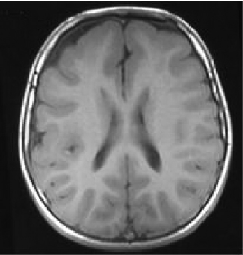
We analyzed the electroclinical findings in two patients with bilateral posterior agyria–pachygyria. Both patients presented with mental retardation, mild motor deficit and epilepsy. The electroclinical findings were characterized by frequent tonic or atonic generalized seizures with occasionally simple or complex partial seizures. Interictal electroencephalography (EEG) showed occipital spikes and diffuse polyspike-wave paroxysms predominantly in the posterior region. Ictal EEG showed diffuse 10–11 Hz activity. Cerebral magnetic resonance imagings (MRIs) showed thickened cortex in the parieto-occipital lobes, bilaterally and symmetrically. The volume of underlying white matter appeared reduced, and the overlying subarachnoid spaces were enlarged. The occipital horns were dilated. These findings were compatible with agyria–pachygyria of the posterior portions of the brain.
In conclusion, in patients with mental retardation, mild motor deficit and epilepsy characterized by tonic or atonic generalized seizures, interictal EEG with diffuse polyspike-wave paroxysms predominantly in posterior region, posterior focal epileptilorm abnormalities and ictal diffuse 10–11 Hz activity, bilateral parieto-occipital agyria–pachygyria should be considered as a possible etiology. Magnetic resonance image is the best neuroradiological study to identify this disorder of cortical development.


